DO NOT INTUBATE JUST FOR SUCTION

A 35 year-old primigravida, with uncomplicated pregnancy, was admitted to the hospital for induction of labor at 41 weeks’ gestation. Ultrasonography suggested the presence of oligohydramnios. Fetal heart rate monitoring disclosed both late and variable decelerations.  Induction of labor with oxytocin was successful. A 3700-g female infant is delivered and covered with meconium. The infant is limp with agonal respirations and a heart rate of 92.


     Meconium aspiration is the inhalation of the first stool of a newborn, a thick, dark-green substance known as meconium. A fetus in distress in utero will often pass meconium in the amniotic fluid, and this is more likely to occur in infants who are term or post term (36->42 weeks gestation). Meconium aspiration usually results in early onset respiratory distress and has several deleterious mechanical, chemical and inflammatory effects on the respiratory system. 1

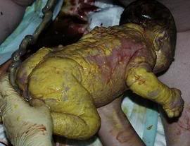
(http://www.mothering.com/forum/306-unassisted-childbirth/1063912-ucing-meconium-aspiration.html)

      A mechanical effect is the obstruction of the airway with meconium, which could lead to atelectasis or hyperinflation. This may alter the ventilation to perfusion ratio or result in barotrauma. Normally, surface tension is reduced by the presence of surfactant. However, infants with meconium aspiration have an increase in surface tension due to the loss of a phospholipid substance, which forms surfactant. This chemical effect of surfactant inhibition mainly results in decrease in lung compliance. In addition, lung injury is caused by the release of the inflammatory mediators that lead to pneumonitis or epithelial airway injury.2

     Research has found that prolonged labor, method of delivery, premature rupture of the membrane, presence of meconium on the infant, fetal distress and fetal asphyxia are all risk factors of developing meconium aspiration and respiratory distress.3 Signs and symptoms of infants with meconium aspiration include cyanosis, nasal flaring, retraction on inspection and grunting. Additionally, auscultation of the lungs may reveal crackles.4

    A variety of modalities and methods can be used in the diagnosis of meconium aspiration. One is through observation of the presence of meconium in the airways by visualizing the vocal cords. Blood gas abnormalities may indicate the development of respiratory distress due to meconium aspiration. Another modality that has played a significant role in the diagnosis of respiratory distress and meconium aspiration is the utilization of chest x-rays .5 Chest x-rays may reveal patchy infiltrates and hyper-inflated lungs, which are commonly seen in infants with meconium aspiration.6

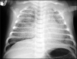
http://www.adhb.govt.nz/newborn/TeachingResources/Radiology/LungParenchyma.htm#RDS


   After the diagnosis of meconium aspiration has been confirmed, it is important to treat the patient as soon as possible to prevent any further complications. The treatment depends on the severity of illness and underlying cause. Suction of the meconium through bulb or suction catheter could be beneficial, but do not intubate the patient just for the sake of suctioning. One of the main treatments used for meconium aspiration is surfactant replacement therapy. Surfactant administration tends to reduce the need for supplemental oxygen, the duration of mechanical ventilation, the incidence of developing pneumothorax and the mortality rate. Another type of therapy used is high frequency oscillatory ventilation (HFOV). However, although it is widely used and has potential benefits, there is a lack of strong evidence supporting the use of HFOV over conventional mechanical ventilation. In very severe cases, infants may develop septic shock or experience cardiac complications. Extracorporeal membrane oxygenation (ECMO) therapy is generally used as a last resort in infants who would die without this form of therapy. Special precautions should be taken when dealing with infants suffering from neurological, nutritional, and musculoskeletal disorders because ECMO could have an effect on increasing morbidity.7

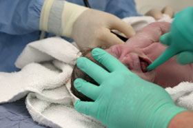
http://www.rosenfeldinjurylawyers.com/meconium-aspiration-syndrome.html


References:

1- Wiedemann, Jeanne R, et al. “Meconium aspiration syndrome.” Neonatal network 27.2 (2008):81-87. Web.


2- van Ierland, Y, and A J J de Beaufort. “Why does meconium cause meconium aspiration syndrome? Current concepts of MAS pathophysiology.” Early human development 85.10 (2009):617-620. Web.


3-Singh, S N, et al. “Respiratory distress including meconium aspiration syndrome in vigorous neonates born through meconium stained amniotic fluid: incidence, onset, severity and predictors at birth.” The Indian Journal of Pediatrics 80.7 (2013):538-543. Web.


4- Edmonds, Patricia. “An introduction to meconium.” Midwifery today with international midwife .111 (2014):32-33. Web.


5- Czarnecki, L M, and Łukasz M Czarnecki. “DIAGNOSTIC IMAGING Assessment of chest X-ray images in newborns with respiratory disorders.” Kardiochirurgia i Torakochirurgia Polska 12.1 (2015):83-86. Web.


6-1. Lynne M, Smith. Meconium Aspiration Imaging.retrived on Friday 04-16-2016 http://emedicine.medscape.com/article/410756-overview


7- Gortner, Ludwig. “Adjunctive therapies for treatment of severe respiratory failure in neonates.” Klinische Pädiatrie 227.2 (2015):51-53. Web.\\\\\\\\

Lung Expansion and Bronchial Hygiene Therapy

Patients with different lung disease and health issues tend to have atelectasis either by breathing lower tidal volume (passive atelectasis) or by mucus plugging (Resorption atelectasis). Different devices are used as a prevention technique to decrease the incidence of atelectasis, which is known as lung expansion therapy. On the other hand, some other devices such as bronchial hygiene therapy devices are used to mobilize the secretions and help the patient to expel these secretions that could be a source of infection.

Lung expansion therapy works under the mechanism of increasing the transpulmonary pressure, which results in increasing lung volumes. An incentive spirometer (IS) is an example of a lung expansion device that is widely used in different situations. It is mostly used with patients who undergo surgery or patients with restrictive lung diseases, who do not breath deeply enough. Special consideration should be given if incentive spirometer is suggested as a therapy because it is not recommended for use with confused or sedated patients since it will not be an effective therapy. Flow or volume oriented devices are different types of IS, but actually, it requires the same maneuver to preform it. Incentive spirometry procedure requires the patient to take a maximal inspiration followed by breath hold to maximize it’s effect. In some situations IS is not effective or it may be contraindicated; in this case, other devices are suggested such as intermittent positive pressure breathing (IPPB). Even though IPPB is not widely used as before it still has a role in lung expansion therapy for carefully selected patients when other lung expansion therapy is not effective. During IPPB therapy, pressure, sensitivity to the patient’s effort to initiate a breath, flow and oxygen percentage are controls that the respiratory therapist would adjust according to the patient’s need. Other lung expansion modalities such as continuous positive pressure (CPAP) or bi-level continuous positive pressure depend on various ways of establishing a positive pressure during exhalation to recruit collapsed alveoli.1


Patients with copious secretions due to diseases that affect the normal cough mechanism, or mucociliary transport system abnormalities are indications for bronchial hygiene therapy. The goal of bronchial hygiene therapy is to remove retained secretions, improve gas exchange and decrease work of breathing. Choosing the appropriate device for the patient depends on the patient’s condition, age and the ability to follow directions. Bronchial hygiene therapy varies between advanced devices that are used in the hospital to simple devices that could be portable or designed for home care use. Cystic fibrosis patients tend to have a large amount of secretions that result in frequent infections. To prevent secretions from blocking their airways, as I mentioned before, it is ideal to use bronchial hygiene therapy. Different modalities are suggested such as chest physiotherapy that is implemented by either percussion or vibration; it also could be combined with postural drainage. Flutter valves or PEP therapy that depend on creating back pressure or vibration to the lungs could be other recommended devices with the advantages of being portable as well as being delivered in conjunction with aerosol therapy. If long periods of therapy were needed, high frequency chest oscillation that has the effect of applying vibration externally to the chest would be a great suggestion for bronchial hygiene therapy. 2


To prevent a patient’s condition from worsening due to breathing at low lung volumes, and to maintain maximal lung function, various kinds of therapy can be used according to the underlying cause to reach therapeutic goals.


Reference:

1-Mosby’s Respiratory care Equipment book

2-Egan’s Fundamentals of Respiratory Care book


Medical gases in Hospitals

The need for using different medical gases in hospitals depends on the patient condition, the underlying disease and the role of the medical gas in treating the disease. Oxygen, nitric oxide, and heliox (the combination of helium and oxygen) are examples of various types of gases that are being uses in the hospital.


Oxygen therapy is the number one drug that is used in hospitals to treat patients with hypoxemia, myocardial infarction, or short-term use for surgeries. It could be implemented on the patient through various delivery devices, which are classified as low and high flow oxygen therapy devices depending on whether the total flow generated by the device is enough to meet the patient inspiratory flow demands. A continuous evaluation of the patient’s condition and response to gas administration is crucially required throughout the treatment. That will give the therapist crucial information about the patient’s improvement or deterioration. In some cases adding oxygen to adjacent therapy will be beneficial. For example, hyperbaric oxygen therapy that is the use of oxygen at a pressure above atmospheric pressure to promote the effect of oxygen. Air embolism as a result of deep diving, and carbon monoxide poisoning from smoke inhalation are indications for hyperbaric therapy.


Mixing helium that has low gas density with the oxygen is effective in treating respiratory conditions associated with high airways resistance such as asthma, bronchitis and chronic obstructive pulmonary disease2. The use of heliox is not limited to the aforementioned diseases; it has been demonstrated in a few studies that it may have a beneficial effect in treating patients with acute respiratory distress syndrome3, in addition to patients with croup. Because of the low density of helium, it decreases turbulent flow in the airways thereby decreasing the work of breathing.2 It is important to know that heliox can be used with both intubated and non-intubated patients. During heliox therapy, it is imperative that the respiratory therapist monitor the patient very closely to prevent hypoxemia and any unwanted complications since the oxygen concentration delivered with heliox therapy may be lower than that required by the patient. Heliox mixtures usually contain either 80% helium and 20% oxygen or 70% helium and 30% oxygen.


It is vital for respiratory therapists to know the concentration of each gas in the different gas mixtures, and their effect, since that will aid in choosing the optimal therapy for the patient. Moreover, respiratory therapists should be able to select the appropriate delivery device that is suitable for the gas mixture, and to to meet the patient’s inspiratory flow demand.


Reference:

1- Mosby’s Respiratory care Equipment book

2- Chevrolet, J C. “Helium oxygen mixtures in the intensive care unit.” Critical care 5.4 (2001):179-81. Web.

3- Yilmaz, Sema, et al. “The effectiveness of heliox in acute respiratory distress syndrome.” Annals of thoracic medicine 8.1 (2013):46-52. Web.

Adrenergic Bronchodilator

Several types of medications are usually used to treat patients with various respiratory diseases. A medication’s mechanism of action depends on the target outcome. Medications are classified as adrenergic, antiadrenergic, cholinergic, anticholinergic and muscarinic. This classification depends on their action in stimulating or blocking the effect of a specific neurotransmitter (e.g., norepinephrine and acetylcholine) on different receptors1.


Adrenergic bronchodilators are widely used by respiratory therapists, to treat patients with diseases that result in decreased airflow or airway obstruction. Patients with asthma, emphysema, bronchitis and cystic fibrosis usually have a better outcome by using adrenergic bronchodilators. For instance, using a short acting Beta-2-adrenergic agent will be ideal with status asthmaticus or patients with acute airway obstruction. Conversely, patients with controlled asthma or chronic obstructive disease tend to use long acting agents. Also, racemic epinephrine is the choice of medication to treat post extubation swelling, croup and epiglottitis. Adrenergic bronchodilators have a stimulation effect on the alpha, beta 1 and beta 2 receptors. They act in increasing heart rate, blood pressure and relaxation of bronchial smooth muscles1.


Even though adrenergic bronchodilators have a great role in treating patients, respiratory therapists should keep in mind special considerations while using these medications. Several side effects may occur with the usage of bronchodilators. Tremor, restlessness, insomnia and weakness are examples of the adverse effects of bronchodilators on the central nervous system. Increased heart rate, palpitation, peripheral vasoconstriction that could result in higher level of blood pressure, bronchial irritation, and edema, are adverse effects of these medications on both the pulmonary and cardiovascular systems 2.


To assess the effectiveness or the adverse effects of bronchodilator treatment, regular patient assessment is needed. Pulmonary function test and peak flow meter would be a useful indicator to monitor the effectiveness of the therapy; that is by assessing the reversibility of the airway obstruction through measuring airflows. Additionally, arterial blood gas results and saturation via pulse oximetry will give a beneficial idea about the adequacy of oxygenation and ventilation in COPD and asthmatic patients1. Moreover, continuous hemodynamic monitoring in addition to the patients’ appearance will reflect the response to the treatment and the patient condition.


Respiratory therapists should be knowledgeable about various medications that are used routinely in the practice of respiratory care, in addition to their adverse effects. This will help them in choosing the appropriate medication for each patient with a specific cardiopulmonary disease.


Reference:

  • -Egan’s Fundamentals of Respiratory Care book.
  • Clinical Practioner’s Poket Guide To Respiratory Care by .Dana Oakes .

Monitoring Devices

Different devices are used by the respiratory therapist to monitor a patient’s respiratory status and to ensure that the therapy is being delivered as intended.

An oxygen analyzer, is a very important device in our profession, it measures the percentage of oxygen being delivered to a patient receiving supplemental oxygen. The sensor of the oxygen analyzer can be connected to the oxygen delivery system proximal to the patient to measure the inspired oxygen concentration. One very common example in the neonatal area is with the use of an oxyhood, where the oxygen analyzer is used to measure the percentage of oxygen being supplied to the baby. Assuring a precise oxygen percentage is critical with neonates as they are more susceptible to the adverse effects of a, high FiO2, which will lead to oxygen toxicity related complications, such as retinopathy of prematurity, bronchopulmonary dysplasia, and respiratory distress syndrome (RDS. On the other hand, if the neonate does not receive enough oxygen, it may cause brain damage due to hypoxia. That is one example of why having a precise measurement of oxygen is critical in health care. 1-2

Oximetry is a simple, noninvasive and relatively inexpensive patient monitor that gives valuable information about the patient’s oxygenation status. Specifically it measures the oxygen saturation of patients. It has a sensor that is placed on the finger or other sites with good capillary blood flow. It works by using a light source on one side of the sensor that sends infrared and red light through the skin to a light detector on the other side of the sensor. Hemoglobin that is saturated with oxygen absorbs more infrared light whereas hemoglobin that is less saturated with oxygen absorbs more red lights. A microprocessor calculates this difference, which is displayed as oxygen saturation. Common sites for placing the pulse oximeter sensor are the fingertips, earlobes, toes, and occasionally, the forehead. Dark skin and nail polish may interfere with light transmission through the skin, resulting in inaccurate measurement. In addition, low perfusion may give inaccurate readings, which we as health care providers should have in mind when treating hypotensive or hypothermic patients who tend to have low perfusion. Another case where the pulse oximeter may have an inaccurate reading is in a patient with carbon monoxide poisoning, which is commonly seen in fire victims. The reading will be affected since carbon monoxide has a great affinity to bind with hemoglobin. In this case, the device will display a falsely high reading for arterial oxygen saturation even though the patient is hypoxic. Consequently, we should not use pulse oximetry in situations such as smoke inhalation where carbon monoxide levels are high, but draw an arterial blood gas and perform co-oximetry. 1-2-3

Dealing with critically ill patients requires the respiratory therapist to be knowledgeable about different devices that help in managing various conditions. Improving his/her critical thinking skills and knowledge regarding the principles of operation of these devices and correctly interpreting the information generated by the various devices are important in helping the respiratory therapist provide safe and effective care.

Reference:

1-Egan’s Fundamentals of Respiratory Care book

2-Mosby’s Respiratory care Equipment book

3-http://www.rtmagazine.com/

Patient Assessment

Each respiratory therapist should possess not only the clinical skills but also, the knowledge and the decision-making skills to diagnose a patient or to monitor the progress of a specific respiratory disease. The respiratory therapist should also understand the effect of various respiratory care diagnostic and therapeutic interventions as well as the devices that are needed for various interventions. For example, general assessment of the patient including vital signs monitoring, would give the practitioner a good but general idea about a patient in respiratory distress, but linking the assessment with additional information such as lab data, pulmonary function tests, and chest radiography will lead to more definitive diagnosis and treatment.

Pulmonary function testing is a way to measure lung volumes and airflow. It is used for the purpose of the diagnosis and measurement of lung impairment. For example, patients with obstructive or restrictive lung disease usually undergo PFT to measure their lung volumes and airflow to provide information about the severity of the disease and the appropriate intervention The benefit of performing pulmonary function testing in patients known to have obstructive lung disease, is to evaluate the severity of the disease and the effectiveness of the therapy1. This depends on performing spirometry with pre and post bronchodilator treatment. If the percentage of change in the volume exhaled in the first second (FEV1) of the forced vital capacity (FVC) maneuver is greater than 12%, it indicates that the patient’s airway obstruction is reversible and that the bronchodilator treatment is effective. An ideal example is an asthmatic patient who performs PFT pre and post bronchodilator2. Additionally, PFT performed prior to surgery could give a clear idea about the risk of postoperative pulmonary complications in patients, who will have surgery under general anesthesia.

Thoracic imaging is another tool that supports the diagnosis of lung diseases. Regular chest X-ray provides an instant thoracic image, but computerized tomography (CT) and magnetic resonance imaging (MRI) require additional time with the benefit of more accuracy. Since thoracic imaging reflects the different densities of various substances (air, fat, soft tissue, and bone) contained in the organs within the thoracic cavity, this will aid diagnosis of a specific disease depending on its characteristics. For instance, pleural effusion, atelectasis or infiltrates tend to appear radiopaque while emphysema or pneumothorax will appear radiolucent. In addition, a chest radiograph serves as a good indicator of the position of artificial airways such as the endotracheal tube. This is important since misplacement of the endotracheal tube could lead to several complications.2-3

Given the diagnostic value of PFTs and chest x-rays, the respiratory therapist should have the ability to interpret the results provided by these diagnostic tools and recommend appropriate interventions.

Reference:

  • Koegelenberg, C. F., Swart, F., Irusen, E. M. (2013). Guideline for office spirometry in adults, 2012. SAMJ. South African medical journal, 103(1), 52-62.
  • Clinical Practioner’s Poket Guide To Respiratory Care by .Dana Oakes .
  • -Egan’s Fundamentals of Respiratory Care book

Effect of the gas laws in the respiratory field

      Understanding the properties of gases, liquids and the various laws regarding them, is important for respiratory therapists, since, those concepts will be merged into their practice. By knowing these concepts, respiratory therapists will have a wide range of knowledge, which will allow them to optimize patient care.

Hyperbaric oxygen therapy depends on the concept of treating various conditions by exposing the patient to pressures above the atmospheric pressure. Different situations such as carbon monoxide poisoning, decompression sickness, smoke inhalation, skin grafts and air embolism could be treated by hyperbaric oxygen therapy.

One of the concepts that hyperbaric oxygen therapy depends on is Boyle’s law. Boyle’s law states that there is an inverse relationship between the pressure exerted by a gas and its volume if the temperature is constant. Due to the high level of pressure in the hyperbaric oxygen therapy there will be a decrease in the volume of the air emboli. This is considered as one of the clinical applications of Boyle’s Law.

Alveolar and arterial partial pressure of oxygen will be increased during hyperbaric therapy. The increase in the alveolar partial pressure of oxygen will be due to Dalton’s law that explains the relationship between the partial pressure of the gas and the total pressure. Additionally, the increased partial pressure of oxygen in the alveoli results in an increase in the amount of oxygen dissolved in the blood, which is a clinical application of Henry’s law.

Work of breathing could be affected during hyperbaric oxygen therapy. Since increasing the pressure results in increasing the density of gas, correspondingly, increasing the density of the gas, will increase the work of breathing. Additionally, temperature should be continuously monitored inside the hyperbaric chamber considering Charles law that the temperature will increase with increase of the pressure.

Some patients could require mechanical ventilation during hyperbaric therapy. Respiratory therapists should closely monitor the patient-ventilator system since the increased density of the gas may significantly increase the resistance, thereby reducing the flow delivered by the ventilator.

Respiratory therapists do not need to memorize all the gas laws, but they need to understand them, since they play a prominent role in different devices and concepts used by respiratory therapists.

Reference:

1-Egan’s Fundamentals of Respiratory Care book

2-Mosby’s Respiratory care Equipment book

Patient care and safety

Patients are the number one priority in hospitals. Consequently, different methods have been implemented in hospitals to facilitate patients’ quick, uneventful recovery and safety. In fact, every health care practitioner should be aware of the policies, procedures, protocols, and standards aimed at achieving patient safety goals.

Why should respiratory therapists (RTs) know about electrical safety? The answer lies in the fact that respiratory therapists, deal with patients in various situations. Moreover, most patient care devices such as ventilators, IV pumps, and various monitoring systems require electricity to operate. Understanding the concept of electricity and how to prevent electrical shocks, which could lead to major disasters, is an important factor for respiratory therapists. For instance, it is important for RTs to know that the severity of electrical shocks will depend mainly on the current flowing through an electrical circuit. Specifically, electrical shock with low current will cause a minor effect such as a tingling sensation in the extremities. On the other hand, an electrical shock with a high current will cause major problems including diaphragm dysfunction and ventricular fibrillation, which eventually lead to death. Electrical shocks can be prevented by ensuring that all devices are connected to outlets that are grounded. This will prevent the current from passing through the body in the event of a short circuit. Also, devices should be checked periodically by qualified personnel .1

Fire is another major problem when it comes to hospital safety. Since oxygen supports combustion, and hospitals are oxygen-rich environments, there is an increased potential for deadly fires. Every RT should be well trained to act efficiently in the event of a fire. RTs must successfully complete education and training programs to master the essential concept of fire safety. One of those concepts is the proper way to use the extinguisher. PASS is an acronym that RTs can use to help them remember how to use a fire extinguisher in the event of a fire (P-Pull the pin , A- Aim the nozzle ,S-S squeeze the handle and sweep the nozzle from side to side at the base of the fire). Moreover, RTs must be conversant with the core plan in the event of a fire emergency. For instance, they must know the sequence of activities that must be performed when responding to a fire, namely, rescue the patient, alert other personnel, contain the fire and evacuate (RACE ) 1. Different evacuation plans are implemented depending on the hospital’s design. Usually, during a hospital fire, horizontal evacuation is done, followed by vertical evacuation if necessary. Fire safety knowledge will help in implementing the action plan in a timely manner.2

Taking care of the patient is another important factor. Ambulation of a patient is one way that RTs can participate in the patient’s recovery process. Although, there are some limitations to ambulation, such as a patient’s level of consciousness, the benefits of early mobility have been demonstrated by several studies. During the ambulation of patients on mechanical ventilators, care must be taken to support the patient and ensure stability of lines, drains, tubes, and the artificial airway. Although ambulation of critically ill patients can be resource intensive, it has important benefits including, preventing bed sores, atelectasis restoring normal body functions, decreasing the length of stay in the hospital, and speeding up the recovery from different infections. 1-3

All respiratory therapists should always keep in mind that their ultimate goal is to optimize patient care, and should engage with the healthcare team in finding the best way to do that.

Reference

History and development of Respiratory care (RC)

Respiratory care is an important specialty in the medical field Even though respiratory care is a new discipline compared to medicine and nursing it is very well established .

The development of this specialty evolved from the ancient times when resuscitation was performed by using a bellows and mouth to mouth breathing to the development of positive pressure ventilation with microprocessors and complex ventilation modes.1 It is important to realize that a lot of scientists have greatly impacted the development of the respiratory care profession. For example, the work of Boyle , Gay -Lussac , Daltons , and Laplace have significantly enhanced understanding of lung mechanics by explaining the relationship between time , pressure , volume and surface tension .

The respiratory care profession began with technicians who were responsible for the oxygen cylinders and the delivery of oxygen by different modalities. However, the respiratory therapist of today is markedly different in that they provide multiple diagnostic and therapeutic interventions to patients, including oxygen , aerosol and positive pressure ventilation. Mechanical ventilation is considered to be one of the revolutions in the history of respiratory care, and has developed from the use of noninvasive negative pressure ventilators such as the iron lung to the use of ventilators capable of delivering different modes of ventilation . 1

The role of the respiratory therapist (RT) is expanding and now includes providing services in different areas such as education, hospital critical care, rehabilitation programs , home care, protocol development, and research ..Respiratory therapists must be willing to participate in team work, commit to continuous learning, and provide quality care. Good leadership skills are also required to be an effective respiratory therapist. Additionally, it is crucial to demonstrate professionalism at work, which involves technical and behavioral competence to assure the provision of high quality care to patients .

The American Association for Respiratory Care (AARC) is the professional organization that promotes the respiratory care profession and advocates for respiratory therapists in the United States. The AARC has a leading role in disseminating knowledge about respiratory care all over the world . The organization assists respiratory therapists with multiple activities to develop their knowledge and skills . Furthermore, it provides continuing education opportunities that help them to maintain their licenses in different states after successful completion of the registered respiratory therapist (RRT) exam administered by the National Board for Respiratory Care (NBRC) . As matter of fact, the NBRC offers t exams that allows RTs to be credentialed as a specialist in different aspects of respiratory care. In addition, the Committee on Accreditation for Respiratory Care ( CoARC) accredits respiratory care programs to enhance the quality of respiratory therapy education . Qualified physicians who are either pulmonologists or anesthesiologists provide medical direction for respiratory therapists working in any healthcare setting, and a select group of physicians known asthe board of medical advisors ( BOMA )2 provide advice related to medical issues to the AARC.

Respiratory care is a profession I would recommend to anyone considering a career in the healthcare field. The profession is expected to grow in the United States because of a projected increase in individuals with respiratory impairment caused by smoking, pollution, and an aging population with chronic conditions3.

Reference :

1- Egan’s Fundamentals of Respiratory Care book

2-WWW.AARC.org

3-http://www.bls.gov/ooh/healthcare/respiratory-therapists.htm